EEG stands for electroencephalography. It measures voltage differences on the surface of the head that are produced, indirectly, by the activity of the brain underneath. It is over a century old, it is the backbone of clinical sleep and epilepsy work, and it is the only widely available method that follows brain activity at the timescale the brain actually operates on.
What the signal is
A single neuron firing produces far too small a field to detect from outside the skull. What EEG picks up is the summed effect of many thousands of neurons — specifically the slower post-synaptic potentials in the dendrites of pyramidal cells in the cortex, which are aligned perpendicular to the surface and so add up rather than cancel out.
Three consequences follow from that, and they explain most of what EEG can and cannot do:
It sees the cortex, not the brain. Structures deeper than the cortical surface contribute very little to a scalp recording. Anything happening in the thalamus, the hippocampus or the brainstem is visible only through its effect on the cortex above it, if at all.
It sees synchrony, not activity. A region working hard but firing out of step produces a smaller signal than a region idling in unison. This is genuinely counter-intuitive and it is the source of a great deal of confused interpretation — a large amplitude does not mean a busy brain.
It is smeared. The signal passes through cerebrospinal fluid, skull and scalp on its way out, and the skull in particular is a poor conductor. The result is spatially blurred: an electrode does not read the patch of cortex directly beneath it so much as a weighted average of a wide area around it.
What is actually recorded
EEG is always a difference. An electrode on its own reads nothing meaningful; every channel is the voltage between one electrode and another, or between one electrode and a reference. Change the reference and every number changes with it, which is why comparing raw amplitudes between two systems that referenced differently is not a comparison at all.
The amplitudes are small. Scalp EEG in an adult is typically in the range of tens of microvolts — millionths of a volt. Mains electricity radiating from the wiring in the wall arrives at the same electrodes at a similar or larger amplitude, which is why a mains notch filter is standard and why a poor electrode contact is so destructive: it is not that the brain signal gets weaker, it is that everything else gets relatively louder.
A number in microvolts only means something if the system was calibrated to produce microvolts. Plenty of consumer devices, ours included at the time of writing, report raw converter counts instead. Counts are perfectly usable for comparing one moment to another on the same device, and meaningless for comparing across devices or people.
What EEG is genuinely good at
Timing. Millisecond resolution is EEG's real claim. Functional MRI has vastly better spatial resolution and is roughly a thousand times slower, because it measures blood flow rather than electrical activity. If the question involves when, EEG is usually the answer.
Sleep. The staging of sleep into light, deep and REM is defined by EEG features — slow waves, sleep spindles, K-complexes — alongside eye movement and muscle tone. This is not an inference from heart rate; it is the actual definition, and it is why polysomnography remains the reference standard.
Rhythms. Oscillations at particular frequencies are the most reproducible thing in the whole field. Alpha rising when the eyes close is the classic demonstration, first published by Hans Berger in 1929, and it still works on almost everyone.
Events. Averaging the response to many repetitions of the same stimulus pulls out an evoked potential — a reliable waveform locked to the event. This underpins hearing tests in newborns, among much else.
What EEG cannot do
It cannot read thoughts, words or images. It cannot localise activity precisely without many electrodes and a good deal of modelling, and even then only to the cortical surface. It cannot tell you what someone is feeling. It cannot diagnose a psychiatric condition — there is no EEG signature that identifies depression, anxiety or ADHD with the reliability a diagnosis would require, despite recurring claims to the contrary.
It is also easily fooled by things that are not the brain at all. Blinks, eye movements, jaw clenching, chewing, heartbeat and the mains supply all appear in a scalp recording, and several of them are far larger than the brain activity underneath. Distinguishing signal from artefact is most of the practical work.
Anything on this site describing what a measurement means is describing an instrument, not a person's health. Ninug is not a medical device: it does not diagnose, treat, monitor or prevent any condition, and nothing here should be used to make a medical decision. If something about your sleep or your health concerns you, that is a conversation for a doctor.
Clinical EEG and consumer EEG are different instruments
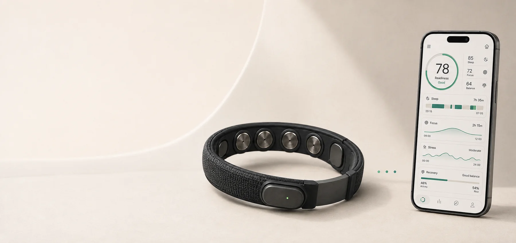
A clinical recording uses many electrodes — commonly 21, often far more — placed by a technician to a standard scheme, with conductive gel and prepared skin, in a room chosen partly for its electrical quiet. A consumer headband has a handful of dry contacts in fixed positions on whatever part of the head a band can comfortably reach, usually the forehead.
The forehead is a compromise, and it is worth being blunt about which way it cuts. It is the most practical place to put a band and the easiest place to get contact without hair in the way. It is also a long way from the back of the head, where the alpha rhythm is strongest, and it sits directly over the frontalis muscle and close to the eyes — so a forehead channel is dominated by blinks and muscle before it is anything else.
That does not make consumer EEG useless. It makes it good at a smaller set of things: eye movement, blinks, muscle tension, gross sleep-wake state, and slow rhythms when the contact is good. It is not a small version of a clinical system.
How to read a claim about EEG
Three questions separate most real claims from most marketing:
Where were the electrodes? A claim about alpha from a forehead-only device deserves more scepticism than the same claim from an occipital electrode, because that is where alpha lives.
What was it compared against? Sleep staging validated against polysomnography is a claim with a referent. Sleep staging validated against nothing is a number with a name on it.
Is the quantity even defined? "Focus", "calm" and "attention" are not EEG measurements. They are interpretations layered on top of one, and the layer is where the assumptions hide.
The rest of this subject
Each of these takes one part of the above and goes further into it.
The signal itself. Delta, theta, alpha, beta and gamma — what the five frequency bands are and what each is honestly associated with. EEG vs ECG — why the two get confused, and why the heart turns up in brain recordings but never the other way round.
The hardware. EEG electrodes — why contact decides everything, and why matching impedance matters more than lowering it. Wearable EEG — what a headband gives up compared with a clinical system, and what it is genuinely better at.
The applications. Sleep, where EEG defines the categories rather than estimating them. Focus, where it does not. Recovery, where it adds something to HRV but less than most products imply. And animal EEG, which is mostly an engineering problem about fur.